Call us now :08045802246

 Send Inquiry
Florescence Trinocular Microscope
Florescence Trinocular Microscope Specification
- Features
- Anti-fungal treated optical parts, ergonomic design, high-stability frame, dust cover included
- View Head
- Trinocular (for simultaneous camera and visual observation)
- Spare Parts
- Replacement bulbs, fuses, filters, eyepieces, and objectives available
- Focus System
- Coaxial coarse & fine focusing mechanism
- Theory
- Florescence observation with trinocular head, suitable for biological and pathological research.
- Drawtube
- Trinocular
- Sensor
- High-resolution optical sensor for image clarity (for camera attachment)
- Resolution
- Up to 1920 x 1080 pixels (depends on camera used)
- Interface
- Standard camera interface compatible with C-mount adapter
- Frame Rate
- 30 FPS (camera dependent)
- Focal Distance
- Adjustable via coaxial coarse and fine focusing knobs
- Magnification
- Up to 1000x with standard objectives
- Dimensions
- Approx. 430 x 260 x 420 mm
- Focus Range
- Coarse (25 mm) and Fine (0.002 mm) adjustment
- Eyepieces
- Wide field 10X/18 mm
- Eyepiece Tube
- Inclined at 45, rotatable 360, for comfortable viewing
- Illumination
- LED or Mercury lamp with brightness adjustment; Blue, Green, Yellow, Ultraviolet filters present
- Coarse Adjustment Range
- 25 mm
- Fine Adjustment Range
- 0.002 mm per division
- Working Stage
- Double layer mechanical stage; 135 x 140 mm; X-Y movement 75 x 50 mm
- Still Image Capture Resolution
- Up to 5MP/8MP/12MP (dependent on camera)
- Video Capture Resolution
- Full HD (depends on camera)
- Image Format
- JPEG, BMP, PNG and video formats via camera
- Interpupillary Distance
- 55-75 mm adjustable
- Objective Achromatic
- Achromatic objectives (4X, 10X, 40X(S), 100X(S, Oil))
- Condenser
- Abbe condenser NA 1.25 with iris diaphragm
- Light Source
- Fluorescence attachment Mercury or LED Lamp (excitation filters included)
- Stage Movement
- Smooth X-Y mechanical movement with position vernier scales
- Arm Stand
- Sturdy arm for vibration-free operation
- Dust Cover
- Protective cover supplied as standard accessory
- Body Material
- Anti-rust, corrosion resistant alloy construction
- Packed Weight
- Approx. 8.5 kg
- Power Supply
- AC 220V/110V 10%, 50/60Hz
- Camera Port
- Dedicated phototube for digital camera connectivity
- Usage
- Ideal for cytology, immunology, histopathology, and microbiology labs
- Filter Cubes
- Blue, Green, Yellow, UV filter holders for fluorescence imaging
- Nosepiece
- Revolving quadruple nosepiece for objective switching
About Florescence Trinocular Microscope
SPECIFICATIONS
Florescence Trinocular Microscope
Epi-Fluorescence Illuminator: The Epi-illumination is through 100W High pressure Mercury Lamp or 5W LED in a lamp housing attached to the fluorescent filter block. The exciting light spectrum range is 350nm-580nm. The Fluorescent Light spectrum range is 420nm-650nm. Two exciting filters (EX) - Blue Light (EX-490) and Green Light (EX-545) color separation prism (DM) comprise of blue light (BA-530), green light (BA-590). Central position provides ordinary transmission light from the base for normal microscopy work. Illumination Box is provided with a power supply of 200V or 110V and HBO, super high pressured spherical mercury vapor lamp 100W. UV protection screen is provided to safeguard the operator from harmful radiations.
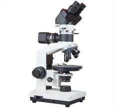
Fluorescent Attachment (Mercury or LED Option)
An ideal attachment for research of biology, tumology, cytology, immunology and Hematology. It is suited for fluorescent Microscopy used in venereal disease examination and immune less diagnosis. At the same time it is also used for analysis of sedimentary rocks, inspection of impurities in semiconductors, environmental protection and micro chemistry.
Superior Fluorescence Imaging
Equipped with filter cubes for blue, green, yellow, and UV wavelengths, this microscope allows detailed fluorescence observation of biological samples. Its built-in fluorescence attachment and high-quality objectives enable clarity, sensitivity, and contrast across a wide range of specimens, making it ideal for tissue and cellular investigations.
Ergonomic and Robust Design
Designed with a sturdy anti-rust frame and smooth stage mechanics, the microscope ensures vibration-free operation and precise X-Y movement. The inclined and rotatable trinocular head allows comfortable viewing and simultaneous camera attachment for documentation, supporting extended periods of research with ergonomic ease.
Versatile Camera and Data Connectivity
A dedicated phototube and standard C-mount interface allow seamless integration of high-resolution cameras. Capture images and Full HD videos in JPEG, BMP, PNG formats, enabling researchers to analyze, save, and share data efficiently for clinical presentations or educational purposes.
FAQs of Florescence Trinocular Microscope:
Q: How does the trinocular microscope facilitate simultaneous visual and digital observation?
A: The trinocular head features a dedicated phototube for digital camera connectivity, allowing simultaneous observation through the eyepieces and image capture or video recording via camera for real-time documentation.Q: What are the primary benefits of using the included filter cubes for fluorescence imaging?
A: Blue, green, yellow, and UV filter cubes enhance the separation and visualization of specific fluorophores, making it possible to study diverse cellular structures or pathogens with high contrast and accuracy during fluorescence experiments.Q: When is it recommended to use the mercury lamp versus LED illumination?
A: The mercury lamp is preferred for applications requiring strong ultraviolet excitation for particular fluorochromes, while the LED provides adjustable, energy-efficient lighting, suitable for general fluorescence imaging and routine observations.Q: Where can the Florescence Trinocular Microscope be deployed for optimal use?
A: This microscope is ideal for cytology, immunology, histopathology, and microbiology laboratories in hospitals, research institutes, universities, and teaching environments where advanced fluorescence techniques are utilized.Q: What process is required for switching between objectives during sample analysis?
A: The revolving quadruple nosepiece allows swift and precise switching between achromatic objectives (4X, 10X, 40X(S), 100X(S, Oil)), facilitating versatile examination of specimens at multiple magnifications without refocusing.Q: How does the microscope support precise sample positioning?
A: The double-layer mechanical stage, equipped with smooth X-Y movement and vernier scales, enables meticulous positioning and tracking of samples, enhancing accuracy in both observation and image documentation.Q: What is the advantage of the anti-fungal treated optical components?
A: Anti-fungal treatments protect optical parts from deterioration in humid environments, ensuring consistent image clarity, longevity of components, and reliable performance in varied laboratory conditions.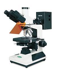
Tell us about your requirement

Price:
Quantity
Select Unit
- 50
- 100
- 200
- 250
- 500
- 1000+
Additional detail
Mobile number
Email

More Products in LABORATORY MICROSCOPE Category
Research Polarizing Microscope
Price 22000 INR / Piece
Minimum Order Quantity : 1 Piece
Working Stage : Rotatable circular stage, 360 graduated, 150 mm diameter, vernier scale
Illumination : Transmitted Koehler illumination (Halogen 6V/20W or LED equivalent)
Theory : Other, Polarized Light Microscopy for optical mineralogy, geology, and material science analysis.
Light Source : Halogen lamp 6V/20W or LED multiple intensity with external power supply
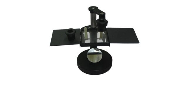
20x Magnification Stereo Dissecting Microscope
Price 900.0 INR / Piece
Minimum Order Quantity : 5 Pieces
Working Stage : Flat circular stage with base illumination Square Yard (yd2)
Illumination : Builtin LED base illumination
Theory : Stereo Microscope
Light Source : Integrated base lighting
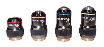
Microscope Objectives
Working Stage : Not applicable for objectives
Illumination : Via microscope light source
Theory : Other, Optical magnification using lenses
Light Source : Not applicable for objectives
Tool Maker Microscope
Working Stage : 130 mm x 125 mm, rotary stage with micrometer slides; XY movement 25 mm in each direction
Illumination : Reflected & transmitted halogen light
Theory : Other, A tool makers microscope is a precision measuring instrument designed for inspection, measurement, and calibration of small parts and tools under magnification.
Light Source : Halogen lamp 6V/20W for illumination
Our Products
- RESEARCH MICROSCOPE
- LABORATORY INSTRUMENTS
- LABORATORY MICROSCOPE
- CHEMISRTY EQUIPMENTS
- OPTICAL BENCHES
- AUTOMATIC PROJECTORS
- HUMAN FIBRE MODELS
- BIOCHEMISTRY EQUIPMENTS
- BIOLOGY LAB EQUIPMENT
- DIGITAL THERMOMETER
- DIGITAL HYDROMETER
- ELECTRONICS ITEMS
- PHYSIOLOGY TESTS
- PHYSIC RESEARCH INSTRUMENTS
- SODA GLASSWARE
- BOROSILICATE GLASSWARE
- PHARMACY EQUIPMENTS & INSTRUMENTS
- Thermometer
- Microscope's Camera
- Microscope's software
- Histopathology
- Operating Microscopes
- Biotechnology Instruments
- Ophthalmic Equipment
- Molecular Biology
- D-Pharmacy Equipment
- Communication Lab Instruments
- Civil Engineering
- Chemistry
- Physics
- tuning fork
- Inverimental
- Hot Air Oven
- Mortuary Chamber

No.-3111/12, Cross Road No. 10, Kacha Bazar, Ambala Cantt - 133001, Haryana, India

Mr Anil Kumar Rana
(Proprietor)
Mobile :08045802246
 Send Inquiry
 Send SMS
Developed and Managed by Infocom Network Private Limited.
