Call us now :08045802246

 Send Inquiry
Trinocular Comparison Microscope
Trinocular Comparison Microscope Specification
- Spare Parts
- Replacement lamps, fuse, eyepieces, objectives available.
- Features
- Simultaneous side-by-side specimen comparison, optional digital documentation, rugged construction.
- View Head
- Trinocular, dual optical path for left and right specimen viewing.
- Focus System
- Coarse and fine coaxial focusing drive.
- Theory
- Used to compare two specimens side by side for forensic, biological, and material analysis.
- Drawtube
- Trinocular head for simultaneous viewing and camera attachment.
- Sensor
- Compatible with digital imaging sensors (with adapter).
- Resolution
- High optical resolution suitable for forensic detail observation.
- Interface
- Standard trinocular port for camera interface.
- Frame Rate
- 30 fps (when paired with digital imaging system).
- Focal Distance
- Adjustable; typically 50mm to infinity depending on objectives.
- Magnification
- 10x to 40x (standard); higher with optional objectives.
- Dimensions
- Approx. 46 x 28 x 45 cm.
- Focus Range
- Coarse and fine focus adjustment, range up to 25mm.
- Eyepieces
- Paired 10x/22mm wide field eyepieces.
- Eyepiece Tube
- Inclined at 45°, rotatable 360°.
- Illumination
- Incident and transmitted halogen lamp illumination, adjustable intensity.
- Coarse Adjustment Range
- Up to 30 mm vertical movement.
- Fine Adjustment Range
- 0.002mm precision fine focus movement.
- Working Stage
- Double plate mechanical comparing stage with slide holders, 75mm x 50mm.
- Still Image Capture Resolution
- Dependent on attached camera; typically 5MP or higher.
- Video Capture Resolution
- Dependent on attached camera; up to 1920x1080 Full HD.
- Image Format
- Supports digital image formats via camera attachment (JPEG, BMP, etc.).
- Interpupillary Distance
- Adjustable from 55 to 75mm.
- Objective Achromatic
- 4x, 10x, 20x, 40x achromatic objectives.
- Condenser
- Abbe condenser, NA 1.25, with iris diaphragm.
- Light Source
- Halogen or LED lamp, brightfield/oblique illumination switchable.
- Camera Mount
- C-mount adapter included for digital camera integration.
- Weight
- Approximately 13 kg.
- Comparator Bridge
- Equipped with a high-quality comparator bridge for seamless image alignment.
- Power Supply
- AC 110V/220V, 50/60Hz.
- Body Material
- Sturdy metal alloy frame with anti-corrosive coating.
- Stage Movement
- X-Y mechanical travel with knobs for precise adjustments.
- Optical System
- Infinity-corrected optical system provides distortion-free imaging.
- Field of View
- Wide field (up to 22 mm) for full sample coverage.
- Polarization
- Polarizing filters available for contrast enhancement.
- Usage
- Ideal for forensic, metal, fiber, and document comparison.
About Trinocular Comparison Microscope
Specifications
Trinocular comparison microscope
Trinocular comparison microscope developed about four decades ago in consultation with the leading Scientists and Criminologists of world repute, thereafter the instruments are being continually updated. Comparison Microscope are the most advanced & versatile instruments invented for comparative Micro study in Forensic Science, geology,
Metallurgy, Mineralogy, Crystallography and Chemical Microscopy. By means of this sophisticated instrument, two micro samples can be studied under one Eye. It combines the latest opto-mechanical developments in light microscopy. This instrument consist of two microscopes & their split images can be viewed simultaneously, with The image from one being on the left side & the other image on the right side. This Microscope can Also compare fibre, hair, bullets, paint fragments under the same lighting conditions & magnification.
The Microscope, having their own illuminations, quadruple revolving nosepieces, coarse and Fine motion knobs, 120mm x 120mm stages, are mounted upon a heavy built-in light base. Leveling Screws are provided to ensure their exact alignment. Transmitted illumination for transparent objects, Incident light for opaque objects and spot lamps for papers are provided for different applications. The comparison bridge brings both the beam paths together and two images of object under Examination, separated by a thin line are observed side by side in the field of view through a Monocular tube.
Standard comparison Microscope are available with Magnification from 20x to 600x having following optical Combination:
Achromatic Objectives : 4x, 10x and 40x SL
Huy. Eyepieces : Wf10x, H15x, H10x micro And H5x cross.
All paired objectives are flat field, strain free and having Anti- reflection coating. The Microscope is supplied with a special vice type Mechanical stages to hold objects like bullets, coins, Industrial materials etc. and another plate to hold Materials like currency notes, cartons, stamps, finger Prints etc.
Total Weight: 45kg.
Size of cases: 46x42x45cm. & 57x41x26 cm.
Optional Accessories: Photo micrographic Attachment Projection Screen Attachment, Semi Plan Objectives 2.5x, 20x, 60x, & Oil Immersion 100x, Spare Eyepieces, USB Camera with Necessary software etc
Precise Side-by-Side Comparison
Utilizing a high-quality comparator bridge and dual optical paths, this microscope is tailored for detailed analysis of two specimens at once. The wide field of view and high-resolution objectives allow experts to detect minute differences or similarities across metals, fibers, documents, and biological samples, ensuring accurate forensic conclusions.
Advanced Imaging Versatility
Infinity-corrected optics ensure clear, distortion-free magnification ranging from 10x to 40x (expandable), complemented by paired 10x/22mm wide field eyepieces. Adjustable halogen/LED illumination and integrated polarization deliver superior contrast for both transmitted and incident lighting setups, facilitating precise examination under varied conditions.
Convenience and Durability
Built with a robust metal alloy frame and anti-corrosive coating, this microscope is engineered for longevity even in demanding laboratory environments. The trinocular head supports both direct viewing and digital imaging with included camera adapters, providing seamless documentation and image analysis for forensic or research purposes.
FAQs of Trinocular Comparison Microscope:
Q: How does the trinocular comparison microscope enable side-by-side analysis of specimens?
A: This microscope features a dual optical path and a high-quality comparator bridge, allowing users to view two specimens simultaneously in the same field. This setup is essential for accurate forensic comparison, letting analysts seamlessly align, examine, and contrast minute sample differences.Q: What types of samples can be examined with this microscope?
A: The instrument is suitable for a broad range of applications, including the comparative analysis of fibers, metals, biological tissues, and documents. Its wide field of view and adjustable magnification make it ideal for forensic investigation, material science, and document verification.Q: When should the polarizing filters be used during observation?
A: Polarizing filters are best utilized when enhanced image contrast or the investigation of birefringent materials is necessary, such as in fiber analysis or distinguishing different metal structures. The filters help suppress glare and clarify subtle details that might otherwise be difficult to discern.Q: Where can digital documentation be performed using this microscope?
A: Digital documentation is supported through the integrated trinocular head and C-mount adapter, which allow connection to most digital cameras. Images and videos can be captured in formats like JPEG or BMP, facilitating easy storage, sharing, and reporting in laboratory or forensic environments.Q: What is the process for adjusting focus and stage position during observation?
A: Precise specimen positioning is achieved using X-Y mechanical stage knobs, which enable smooth movement. Coarse and fine coaxial focus controls allow both rapid and micro-adjustments, with fine focus precision up to 0.002mm, ensuring critical details are always in sharp view.Q: How does the infinity-corrected optical system benefit users?
A: The infinity-corrected optical design provides distortion-free images across the entire field, allowing for accurate, high-resolution observation regardless of sample thickness. This is crucial in forensic work where clarity and measurement accuracy are paramount.Q: What maintenance and spare parts are available for this microscope?
A: To ensure longevity and consistent performance, replacement parts such as lamps, fuses, eyepieces, and objectives are available through the manufacturer or supplier, supporting ongoing laboratory use with minimal downtime.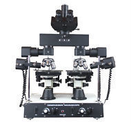
Tell us about your requirement

Price:
Quantity
Select Unit
- 50
- 100
- 200
- 250
- 500
- 1000+
Additional detail
Mobile number
Email

More Products in LABORATORY MICROSCOPE Category
20x Magnification Stereo Dissecting Microscope
Price 900.0 INR / Piece
Minimum Order Quantity : 5 Pieces
Eyepieces : Included (dual eyepieces visible)
Magnification : 20x
Dimensions : Approx. standard microscope size (compact) Meter (m)
Theory : Stereo Microscope
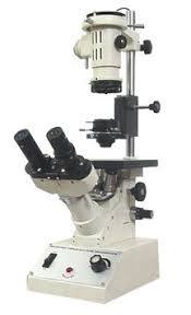
Trinocular Inverted Tissue Culture Microscope
Eyepieces : Paired WF10X/18mm (Wide Field)
Magnification : 40X to 400X (standard with 10X eyepiece and 4X, 10X, 20X, 40X objectives)
Dimensions : 460 x 240 x 530 mm (approx.)
Theory : Other, Inverted tissue culture microscope designed for observing living cells and tissues in culture, suitable for research and clinical laboratories.

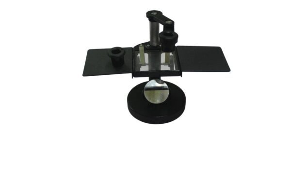
Filler Micrometer Eyepiece
Eyepieces : Single optical eyepiece with integrated micrometer scale
Magnification : 10x (eyepiece) with direct scale view
Dimensions : Eyepiece diameter: 23.2 mm; length: approximately 50 mm
Theory : Other, A filler micrometer eyepiece is an optical device used in microscopes to measure small distances or diameters with high precision, integrating a calibrated scale within the eyepiece for direct measurement.
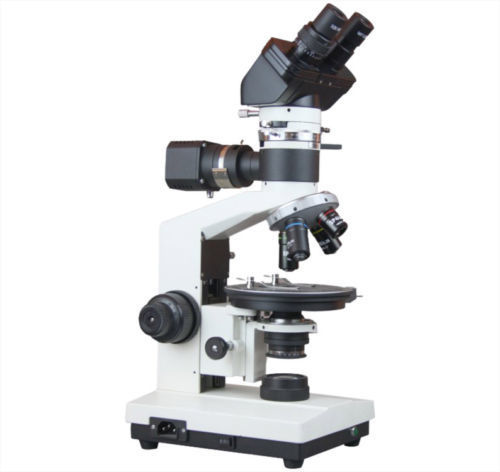
Research Polarizing Microscope
Price 29000.0 INR / Piece
Minimum Order Quantity : 1 Piece
Eyepieces : Widefield, strainfree eyepieces with a 10x or higher magnification.
Magnification : 400x
Dimensions : 220X264X240 Millimeter (mm)
Theory : Other
Our Products
- RESEARCH MICROSCOPE
- LABORATORY INSTRUMENTS
- LABORATORY MICROSCOPE
- CHEMISRTY EQUIPMENTS
- OPTICAL BENCHES
- AUTOMATIC PROJECTORS
- HUMAN FIBRE MODELS
- BIOCHEMISTRY EQUIPMENTS
- BIOLOGY LAB EQUIPMENT
- DIGITAL THERMOMETER
- DIGITAL HYDROMETER
- ELECTRONICS ITEMS
- PHYSIOLOGY TESTS
- PHYSIC RESEARCH INSTRUMENTS
- SODA GLASSWARE
- BOROSILICATE GLASSWARE
- PHARMACY EQUIPMENTS & INSTRUMENTS
- Thermometer
- Microscope's Camera
- Microscope's software
- Histopathology
- Operating Microscopes
- Biotechnology Instruments
- Ophthalmic Equipment
- Molecular Biology
- D-Pharmacy Equipment
- Communication Lab Instruments
- Civil Engineering
- Chemistry
- Physics
- tuning fork
- Inverimental
- Hot Air Oven
- Mortuary Chamber

No.-3111/12, Cross Road No. 10, Kacha Bazar, Ambala Cantt - 133001, Haryana, India

Mr Anil Kumar Rana
(Proprietor)
Mobile :08045802246
 Send Inquiry
 Send SMS
Developed and Managed by Infocom Network Private Limited.
