Call us now :08045802246

 Send Inquiry
Dissecting Microscope
Price 900 INR/ Unit
MOQ : 1 Unit
Dissecting Microscope Specification
- Features
- Heavy stable base, sturdy stand, simple to use, portable, suitable for student biology laboratories.
- View Head
- Monocular vertical view head.
- Spare Parts
- Replacement eyepieces, objectives, mirror, and stage clips available.
- Focus System
- Coarse rack and pinion focusing mechanism.
- Theory
- A Dissecting Microscope is an optical microscope designed for observing low magnification samples using incident light or reflected illumination from above the specimen. It is commonly used for biological studies, entomology, and dissection tasks.
- Drawtube
- Upright, single draw tube with focusing arrangement.
- Sensor
- Not Applicable (Optical viewing only)
- Resolution
- Dependent on magnification; provides clear detail at low magnification (up to 40x).
- Interface
- Manual operation, no digital interface.
- Frame Rate
- Not applicable (no video or digital imaging).
- Focal Distance
- Approximately 50 mm to 150 mm, adjustable via focusing knob.
- Magnification
- Typically ranges from 10x to 40x using combinations of eyepieces and objectives.
- Dimensions
- Approx. 200 mm 150 mm 340 mm (L x W x H).
- Focus Range
- Coarse focusing with adjustable knob, range up to 50 mm.
- Eyepieces
- Wide field 10x (paired); can be fitted with 5x or 15x eyepieces.
- Eyepiece Tube
- Single monocular vertical tube.
- Illumination
- Plano-concave mirror for natural reflected light, with provision for substitute artificial light.
- Coarse Adjustment Range
- Up to 50 mm vertical movement.
- Fine Adjustment Range
- Not available, only coarse adjustment present.
- Working Stage
- Glass plate with two spring clips for holding specimens, size approx 100 mm diameter.
- Still Image Capture Resolution
- Not applicable.
- Video Capture Resolution
- Not applicable.
- Image Format
- Optical image (no digital capture).
- Interpupillary Distance
- Not adjustable (fixed, monocular tube).
- Objective Achromatic
- One pair of achromatic objectives; standard 2x and 4x.
- Condenser
- Not applicable; uses incident or reflected light.
- Light Source
- Reflective plano-concave mirror; external lamp optional.
- Weight
- Approximately 2.5 kg.
- Brand Compatibility
- Compatible with standard and replacement parts from most educational microscope brands.
- Objective Mount
- Revolving arm facilitates quick change of objectives.
- Color
- Ivory white with metallic accents.
- Stage Plate
- Frosted glass plate included for transmitted light observations.
- Mirror Adjustment
- Movable mirror for adjusting incident and reflected light angles.
- Usage/Application
- Ideal for gross dissection, entomological and elementary laboratory work.
- Construction Material
- Sturdy metallic body with enamel finish.
- Base Type
- Horseshoe shaped base for stability.
About Dissecting Microscope
Dissecting Microscope
Square stage with two clips for holding slide clear glass stage matted plate and hand rests 10x and 20x magnifying eyepieces focusing by rack & pinion mechanism plane and concave mirror two hinged joint arm made of brass for magnifier thermacol packing ,dust cover
Microscopes are designed with outstanding technological expertise of which extend the capabilities for education and research work. The quality assurance throughout the manufacturing process ensures demanding performance standards which are matched with exceptional economy in cost. Extra clarity and contrast is provided through our new high performance color corrected infinity optical system. Optical components in the viewing head and objectives are protected with an anti mould system to ensure proper performance in unusually hot and humid environment.
Sturdy Construction for Reliable Use
Built with a durable metallic body and finished with enamel, this dissecting microscope is designed for everyday use in laboratories and classrooms. Its horseshoe-shaped base grants essential stability, providing secure operation while examining specimens. The sturdy structure ensures longevity even with frequent handling.
Precise Observation and Easy Operation
The single, upright monocular draw tube, combined with wide-field eyepieces (10x, replaceable with 5x or 15x), delivers clear, magnified views of specimens. The microscope allows vertical movement up to 50 mm for coarse focusing, while interchangeable achromatic objectives and a movable mirror enable flexible illumination and rapid adjustment to different samples.
Versatile Illumination Methods
This model supports both incident (reflected) and transmitted light observation. The plano-concave mirror can be adjusted to direct natural or artificial light as needed, and the frosted glass stage plate allows for examination of translucent specimens. External lamps can be used when required, providing consistent lighting conditions for detailed studies.
FAQ's of Dissecting Microscope:
Q: How do I use the dissecting microscope for specimen observation?
A: Place your specimen on the frosted glass stage plate, secure it with the spring clips, adjust the mirrored light source to optimize illumination, and use the coarse focusing knob to obtain a clear view through the monocular eyepiece.Q: What are the main applications of this dissecting microscope?
A: This microscope is primarily used for gross dissection, entomological studies, and elementary laboratory work, offering clear views of low-magnification samples such as plants, insects, and small anatomical dissections.Q: When should I adjust the mirror, and how does it affect imaging?
A: Adjust the mirror when changing between natural and artificial light sources or altering the angle of illumination. Proper mirror positioning enhances visibility and contrast depending on the nature of your specimen and the surrounding lighting conditions.Q: Where is this microscope best suited for use?
A: The microscope is well-suited for student biology laboratories, educational institutions, entomological labs, and settings where routine low-magnification observation is needed. Its portability makes it adaptable to various learning environments.Q: What maintenance or replacement parts are available for this microscope?
A: Replacement eyepieces, objectives, mirrors, and stage clips are available for this model. Regular housekeeping-such as cleaning lenses and the mirror, and gently handling all mechanical parts-will ensure longevity and consistent performance.Q: How does the focusing system work on this microscope?
A: The focusing system operates through a coarse rack and pinion mechanism, allowing vertical movement of the microscope head up to 50 mm. This enables easy adjustment to achieve the desired focal distance for sharp, clear images of your sample.Q: What are the benefits of using a monocular dissecting microscope like this?
A: A monocular dissecting microscope is simple to operate, cost-effective, and suitable for educational purposes. It provides clear, wide-field views at low magnification levels, making it ideal for scanning and inspecting larger specimens during hands-on laboratory exercises.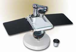
Tell us about your requirement

Price:
Quantity
Select Unit
- 50
- 100
- 200
- 250
- 500
- 1000+
Additional detail
Mobile number
Email

More Products in LABORATORY MICROSCOPE Category
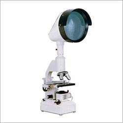
Projection Microscope
Price 12500 INR
Minimum Order Quantity : 1 Unit
Illumination : 6V 20W Halogen lamp
Dimensions : 320 mm x 220 mm x 430 mm
Light Source : Halogen lamp
Magnification : 40x to 1500x
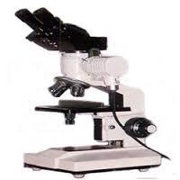
Binocular Metallurgical Microscope
Price 6000 INR / Piece
Minimum Order Quantity : 1 Piece
Illumination : LED illumination adjustable brightness
Dimensions : 340mm x 230mm x 420mm
Light Source : Halogen lamp
Magnification : 40x to 1000x
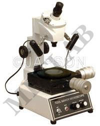
Tool Maker Microscope
Illumination : Reflected & transmitted halogen light
Dimensions : Approx. 350 mm (H) x 200 mm (W) x 300 mm (D)
Light Source : Halogen lamp 6V/20W for illumination
Magnification : Up to 50X (with combination of eyepiece and objective lens)
Microscopelamp
Illumination : Halogen / LED Lamp
Dimensions : Compact design
Light Source : Halogen / LED Bulb
Our Products
- RESEARCH MICROSCOPE
- LABORATORY INSTRUMENTS
- LABORATORY MICROSCOPE
- CHEMISRTY EQUIPMENTS
- OPTICAL BENCHES
- AUTOMATIC PROJECTORS
- HUMAN FIBRE MODELS
- BIOCHEMISTRY EQUIPMENTS
- BIOLOGY LAB EQUIPMENT
- DIGITAL THERMOMETER
- DIGITAL HYDROMETER
- ELECTRONICS ITEMS
- PHYSIOLOGY TESTS
- PHYSIC RESEARCH INSTRUMENTS
- SODA GLASSWARE
- BOROSILICATE GLASSWARE
- PHARMACY EQUIPMENTS & INSTRUMENTS
- Thermometer
- Microscope's Camera
- Microscope's software
- Histopathology
- Operating Microscopes
- Biotechnology Instruments
- Ophthalmic Equipment
- Molecular Biology
- D-Pharmacy Equipment
- Communication Lab Instruments
- Civil Engineering
- Chemistry
- Physics
- tuning fork
- Inverimental
- Hot Air Oven
- Mortuary Chamber

No.-3111/12, Cross Road No. 10, Kacha Bazar, Ambala Cantt - 133001, Haryana, India

Mr Anil Kumar Rana
(Proprietor)
Mobile :08045802246
 Send Inquiry
 Send SMS
 Call Me Free
Developed and Managed by Infocom Network Private Limited.
