Call us now :08045802246

 Send Inquiry
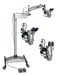
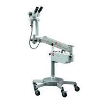
Neuro Surgery Operating Microscope
Price 11000.0 INR/ Piece
MOQ : 1 Piece
Neuro Surgery Operating Microscope Specification
- Power
- 220-240 Volt (v)
- Material
- STAINLESS STEEL
- Weight
- 75 Kilograms (kg)
- Voltage
- 220-240 Volt (v)
- Operate Method
- MANUAL
- Magnification
- 2X-20X
- Focus Range
- 50 Millimeter (mm)
- Eyepieces
- 12.5X
- Eyepiece Tube
- BINOCULAR TUBE
- Illumination
- COXIAL
- Interpupillary Distance
- 55-75 Millimeter (mm)
- Light Source
- LED
Neuro Surgery Operating Microscope Trade Information
- Minimum Order Quantity
- 1 Piece
- Supply Ability
- 1 Piece Per Month
- Delivery Time
- 1 Week
- Main Domestic Market
- All India
- Certifications
- CE CERTIFIED CO. NSIC MSME ISO 9001:2015
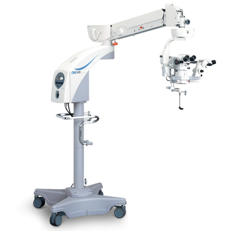
About Neuro Surgery Operating Microscope
Neuro Surgery Operating Microscope
Neurosurgery microscope specifications includea motorized, coaxial illumination system with a high-intensity light source (100,000-150,000 lux) for exceptional brightness, magnification ratios (e.g., 1:6), adjustable optical components like a tiltable binocular tube and objective lenses with focal lengths of 200-350mm, and advanced X-Y translation systems for precise positioning, alongside options for integrated cameras and augmented reality (AR) features to enhance digital visualization and real-time data display.
Key Components and Specifications
-
- Light Source:High-power sources like LED or Xenon lamps are common, with intensities often reaching 100,000-150,000 lux.
- Coaxial Illumination:Ensures the light beam is directly aligned with the optical path for shadow-free, high-contrast viewing of deep surgical fields.
- Filters:Red-free and yellow filters are used to improve tissue contrast, and small spot filters for focused illumination.
- Light Source:High-power sources like LED or Xenon lamps are common, with intensities often reaching 100,000-150,000 lux.
-
- Binocular Tube:A tiltable binocular tube, often adjustable from 0 to 90 or 180 degrees, allows for surgeon comfort.
- Eyepieces:Standard eyepieces are 10x or 12.5x magnification.
- Objective Lenses:Varying focal lengths (e.g., 200mm, 300mm, 350mm) provide different working distances and magnification ranges.
- Magnification:Typically ranges from 2x to 20x, with motorized zoom systems offering ratios up to 1:6 for continuous magnification control.
- Binocular Tube:A tiltable binocular tube, often adjustable from 0 to 90 or 180 degrees, allows for surgeon comfort.
-
- X-Y Translator:A motorized system, often controlled by a foot switch, allows for precise movement of the microscope over the surgical field.
- Focusing:Can be manual or motorized, with standard focusing distances around 50mm.
- Vertical Movement:A broad range of vertical movement, such as 600mm, allows for positioning at various heights.
- X-Y Translator:A motorized system, often controlled by a foot switch, allows for precise movement of the microscope over the surgical field.
-
- CCD Camera Systems:Integrated systems with beam splitters and adapters enable image capture and display for documentation, training, and remote viewing.
- Augmented Reality (AR):Advanced microscopes incorporate AR to display real-time data, such asblood flow visualization,directly within the surgeons field of view.
- CCD Camera Systems:Integrated systems with beam splitters and adapters enable image capture and display for documentation, training, and remote viewing.
Superior Magnification and Precision
With a flexible magnification range from 2X to 20X, the microscope enables surgeons to seamlessly adjust views for detailed observation. The built-in focusing range of 50 mm further enhances the ability to visualize even the most delicate structures in neurosurgery.
Ergonomic Design for Comfort
The binocular eyepiece tube and adjustable interpupillary distance (55-75 mm) allow surgeons of varying preferences to achieve comfortable viewing. Paired with lightweight 12.5X eyepieces and balanced at 75 kg, the instrument supports fatigue-free operation during long procedures.
Advanced LED Illumination for Clarity
Equipped with an efficient coaxial LED light source, the operating microscope delivers crisp, shadow-free illumination. This ensures that deep and narrow surgical fields are adequately lit, which is essential for precision in neurosurgical interventions.
FAQs of Neuro Surgery Operating Microscope:
Q: How do I operate the Neuro Surgery Operating Microscope manually during procedures?
A: The microscope is equipped with easy-to-use manual controls, allowing users to adjust magnification between 2X and 20X, set the focus within a 50 mm range, and modify the interpupillary distance from 55 to 75 mm. Adjustments can be made smoothly to maintain optimal imagery throughout your procedure.Q: What are the main benefits of using this microscope in neurosurgery?
A: This microscope offers high precision through adjustable magnification and coaxial LED illumination, enhancing visibility of neural structures. Its ergonomic binocular design and stainless steel construction ensure durability and comfort, supporting complex surgeries with less fatigue and better outcomes.Q: When is this microscope most effectively utilized?
A: The microscope is ideal for neurosurgical procedures that require detailed visualization of intricate neural tissues, such as tumor removal or aneurysm clipping. Its superior optics and illumination are best used during operations demanding high magnification and consistent clarity.Q: Where can this neuro surgery operating microscope be supplied or exported from?
A: This microscope is manufactured and exported from India. It is available to medical institutions globally through established exporter, manufacturer, and supplier channels, ensuring reliable delivery and support.Q: What is the process for setting up and using the microscope in the operating room?
A: Begin by securely positioning the microscope and connecting it to a power source of 220-240 Volts. Adjust the eyepieces to 12.5X, set the interpupillary distance, and calibrate magnification and focus as required. Activate the LED illumination and verify all fields are clear before the surgical procedure begins.Q: How does the LED coaxial illumination improve neurosurgery outcomes?
A: The LED coaxial illumination provides bright, shadow-free light directly aligned with the optical path. This innovative lighting method enhances the ability to discern minute neural details and ensures accurate depth perception within the surgical field.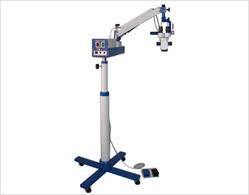

Tell us about your requirement

Price:
Quantity
Select Unit
- 50
- 100
- 200
- 250
- 500
- 1000+
Additional detail
Mobile number
Email

More Products in Operating Microscopes Category

Operating Microscope
Price 12000 INR / Piece
Minimum Order Quantity : 1 Piece
View Head : Binocular head, 360 rotatable
Drawtube : Binocular
Video Capture Resolution : 1920 x 1080 pixels
Image Format : JPG, BMP, AVI
Operating Microscope(Dental)
Price 11500 INR / Piece
Minimum Order Quantity : 1 Piece
View Head : Binocular, inclinable and rotatable.
Drawtube : Other, Binocular, inclined at 45 for improved ergonomics during procedures.
Video Capture Resolution : 1920 x 1080 @ 30 fps.
Image Format : JPEG (still images), MP4 (video via camera module).
Multiple Microscope
Price 11000 INR / Inch
Minimum Order Quantity : 1 Piece
View Head : Multiple viewing attachment for 2, 3, 5, or more users
Drawtube : Other, Binocular & Trinocular available
Video Capture Resolution : Full HD 1080p (as per model)
Image Format : JPEG, BMP (with cameraequipped models)

Operating Microscope
Price 11500 INR / Piece
Minimum Order Quantity : 1 Piece
View Head : Binocular Head with Adjustable Interpupillary Distance
Drawtube : Other, Not available
Video Capture Resolution : Not available
Image Format : Not available
Our Products
- RESEARCH MICROSCOPE
- LABORATORY INSTRUMENTS
- LABORATORY MICROSCOPE
- CHEMISRTY EQUIPMENTS
- OPTICAL BENCHES
- AUTOMATIC PROJECTORS
- HUMAN FIBRE MODELS
- BIOCHEMISTRY EQUIPMENTS
- BIOLOGY LAB EQUIPMENT
- DIGITAL THERMOMETER
- DIGITAL HYDROMETER
- ELECTRONICS ITEMS
- PHYSIOLOGY TESTS
- PHYSIC RESEARCH INSTRUMENTS
- SODA GLASSWARE
- BOROSILICATE GLASSWARE
- PHARMACY EQUIPMENTS & INSTRUMENTS
- Thermometer
- Microscope's Camera
- Microscope's software
- Histopathology
- Operating Microscopes
- Biotechnology Instruments
- Ophthalmic Equipment
- Molecular Biology
- D-Pharmacy Equipment
- Communication Lab Instruments
- Civil Engineering
- Chemistry
- Physics
- tuning fork
- Inverimental
- Hot Air Oven
- Mortuary Chamber

No.-3111/12, Cross Road No. 10, Kacha Bazar, Ambala Cantt - 133001, Haryana, India

Mr Anil Kumar Rana
(Proprietor)
Mobile :08045802246
 Send Inquiry
 Send SMS
 Call Me Free
Developed and Managed by Infocom Network Private Limited.
