Call us now :08045802246

 Send Inquiry
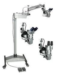
Operating Microscope
Price 12000 INR/ Piece
MOQ : 1 Piece
Operating Microscope Specification
- Focus System
- Motorized/manual high-precision focusing
- Spare Parts
- Supplied with dust cover, spare bulb, and fuse
- Features
- Apochromatic optics, anti-reflective coating, ergonomic design for long procedures, smooth movement, compact footprint, high depth of field
- View Head
- Binocular head, 360 rotatable
- Theory
- Operating Microscope is used for performing microsurgery and offers high precision visual clarity for medical professionals.
- Drawtube
- Binocular
- Sensor
- High-quality CMOS sensor integrated for superior image acquisition.
- Resolution
- Full HD 1920 x 1080 pixels
- Interface
- HDMI, USB 2.0, VGA
- Frame Rate
- 30 fps (Full HD recording)
- Focal Distance
- F=200 mm (fixed)
- Magnification
- Variable, 4x-40x (with multiple objective and eyepiece combinations)
- Dimensions
- Main body: 380 x 345 x 700 mm
- Focus Range
- Manual, coarse and fine adjustment 50 mm
- Eyepieces
- 10x widefield, paired
- Eyepiece Tube
- Inclined 45, rotatable
- Illumination
- LED / Halogen coaxial illumination, fiber-optic, 40,000 Lux
- Coarse Adjustment Range
- 50 mm
- Fine Adjustment Range
- 2 mm
- Working Stage
- Tiltable and height adjustable stage with patient support arms
- Still Image Capture Resolution
- 1920 x 1080 pixels
- Video Capture Resolution
- 1920 x 1080 pixels
- Image Format
- JPG, BMP, AVI
- Interpupillary Distance
- 55-75 mm adjustable
- Objective Achromatic
- f=200 mm achromatic objective standard (optional: f=250 mm, f=300 mm)
- Condenser
- Built-in condenser lens for focused illumination
- Light Source
- LED / Halogen, 12V/100W, long-life bulb
- Power Supply
- 220V/50Hz (standard), 110V/60Hz available
- Packaging
- Shock proof packing with foam lining for safe transport
- Camera Integration
- Integrated digital camera with real-time image transfer
- Weight
- Approx. 28 kg
- Safety Standards
- CE certified, conforms to medical device standards
- Mounting Type
- Mobile stand with castors and locking brakes
- Operating Temperature
- 10C to 35C
- Sterilization Capability
- Removable components designed for autoclave sterilization
- Arm Movement
- Multi-jointed, counterbalanced arm with up/down and lateral movement
About Operating Microscope
SPECIFICATIONS
Plastic Surgical Operating Microscope offered find application in conducting different types of surgeries. These include nose, ear and throat surgery, ophthalmic eye surgery, neurosurgery and others. With the microscopes designed to provide excellent working support in involved procedures, these deliver superior accuracy in working and can be made available with different microscope sections including choice of Optical Heads, Eye Pieces, Eye Inclination, Inter-papillary Distance. We can also offer these in different power choices as per the specific work application needs.
Features:
- Precision functioning plastic surgical operating microscope
- Available with option of manual focusing and positioning
- 320 degree rotatable counter balanced gas springs loaded arm
- Allows easy variable angle viewing
Specification
- Binocular Tube : Ergonomic tilt able viewing 0° - 210° with converging optics
- Magnification : 0.4x, 0.63x, 1.0x, 1.6x & 25x with five step magni changer
- Eye Pieces : WF 10x/16mm paired with eye guards
- IPD Range : 55 - 75 mm by knob
- Working Distance : F=200mm Objective Lens
- Fine Focusing : Motorized with Foot Control
- Light Source : 50W LED up to 60,000 hours of LED lamp life
- Built in Filters : Green & Yellow
- Vertical Movement of Arm : 600mm
- Microscope Carrier : 120° Plastic carrier
Superior Imaging & Integration
This microscope boasts a high-quality CMOS sensor, delivering Full HD images at 1920 x 1080 pixels, with support for still images in JPG or BMP and videos in AVI format. Real-time image transfer via HDMI, USB 2.0, and VGA interfaces allows for easy sharing, documentation, and live collaboration during procedures.
Ergonomic & Mobile Design
The system features a counterbalanced, multi-jointed arm for effortless movement, along with a compact, mobile stand equipped with locking castors for stability. Its design allows for prolonged, comfortable use thanks to an inclined, rotatable binocular head, high depth of field, and anti-reflective coatings.
Effective Illumination & Focus
Achieve precise visualization with fiber-optic LED or halogen coaxial illumination up to 40,000 Lux. The microscope includes both manual coarse and fine focusing (motorized/manual support), with a 50-mm adjustment range and adjustable stage for patient support and alignment.
Reliable & Safe for Clinical Use
Compliant with CE and other medical device standards, the microscope includes autoclave-ready removable parts, shock-proof packaging for safe transport, and is supplied with useful spares. Adjustable power options, a robust build, and an easy-to-clean design make it ideal for demanding clinical environments.
FAQs of Operating Microscope:
Q: How is the integrated camera used during microsurgical procedures?
A: The built-in high-resolution CMOS camera enables surgeons and clinicians to capture Full HD images and videos during procedures. Real-time image transfer via HDMI, USB, or VGA allows for immediate review, documentation, teaching, or remote collaboration without interrupting the surgery.Q: What are the sterilization procedures for the microscopes removable components?
A: All removable components are designed for autoclave sterilization. After detaching these parts, they can be safely processed following standard clinical sterilization protocols, helping maintain a sterile surgical environment.Q: When should I use manual versus motorized focusing?
A: Manual focusing is ideal for situations that require quick, fine-tuned adjustments, while motorized focusing provides high-precision control, especially helpful during lengthy or intricate procedures where hands-free adjustment is beneficial.Q: Where can the operating microscope be installed and operated?
A: Thanks to its mobile stand with locking castors and compact footprint, the microscope is versatile for use in various clinical and surgical settings, such as operating rooms, outpatient clinics, or teaching laboratories.Q: What is the benefit of using an apochromatic objective with anti-reflective coating?
A: Apochromatic optics with anti-reflective coatings enhance image clarity and color fidelity, reducing chromatic and spherical aberrations. This results in highly accurate, sharp visuals, crucial for precision in microsurgical procedures.Q: How does the microscope support documentation and workflow?
A: The integrated camera, multiple interfaces, and high-resolution image capabilities streamline the documentation process, allowing for effortless capture, storage, and transfer of visual data to medical records or external devices.Q: What safety standards does the microscope conform to?
A: The microscope is CE certified and conforms to recognized medical device safety standards, ensuring safe operation, reliability, and suitability for medical environments.
Tell us about your requirement

Price:
Quantity
Select Unit
- 50
- 100
- 200
- 250
- 500
- 1000+
Additional detail
Mobile number
Email

More Products in Operating Microscopes Category

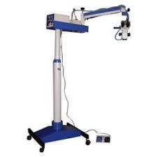
Operating Microscope
Price 11500 INR / Piece
Minimum Order Quantity : 1 Piece
Light Source : LED Light
Focus Range : Not available
Eyepiece Tube : Not available
Eyepieces : Not available
Operating Microscope
Light Source : LED with long operating life
Focus Range : 50 mm 80 mm
Eyepiece Tube : Inclined at 45 for comfortable viewing
Eyepieces : Widefield 10x/16x

Operating Microscope
Light Source : 3W LED lamp (cool illumination), longlasting and energy saving.
Focus Range : Manual coarse & fine adjustment; 50mm coarse, 2mm fine.
Eyepiece Tube : Inclined binocular tube at 45, rotatable 360.
Eyepieces : Wide field eyepiece WF10x/22mm (pair); optional WF16x available.
Operating Microscope
Light Source : Builtin Halogen or LED (configurable)
Focus Range : Manual coarse and fine focusing, 50 mm total travel
Eyepiece Tube : Inclined, 45 degrees for ergonomic viewing
Eyepieces : 10x wide field (paired), diopter adjustable
Our Products
- RESEARCH MICROSCOPE
- LABORATORY INSTRUMENTS
- LABORATORY MICROSCOPE
- CHEMISRTY EQUIPMENTS
- OPTICAL BENCHES
- AUTOMATIC PROJECTORS
- HUMAN FIBRE MODELS
- BIOCHEMISTRY EQUIPMENTS
- BIOLOGY LAB EQUIPMENT
- DIGITAL THERMOMETER
- DIGITAL HYDROMETER
- ELECTRONICS ITEMS
- PHYSIOLOGY TESTS
- PHYSIC RESEARCH INSTRUMENTS
- SODA GLASSWARE
- BOROSILICATE GLASSWARE
- PHARMACY EQUIPMENTS & INSTRUMENTS
- Thermometer
- Microscope's Camera
- Microscope's software
- Histopathology
- Operating Microscopes
- Biotechnology Instruments
- Ophthalmic Equipment
- Molecular Biology
- D-Pharmacy Equipment
- Communication Lab Instruments
- Civil Engineering
- Chemistry
- Physics
- tuning fork
- Inverimental
- Hot Air Oven
- Mortuary Chamber

No.-3111/12, Cross Road No. 10, Kacha Bazar, Ambala Cantt - 133001, Haryana, India

Mr Anil Kumar Rana
(Proprietor)
Mobile :08045802246
 Send Inquiry
 Send SMS
Developed and Managed by Infocom Network Private Limited.
